REVIEW

Alternatives to antibiotics: phage lytic enzymes and phage therapy

About authors

Belozersky Institute of Physico-Chemical Biology,
Lomonosov Moscow State University, Moscow, Russia

Correspondence should be addressed: Pavel Nazarov
ul. Narimanovskaya 22, k,3, kv. 294, Moscow, 117997; moc.liamg@apvorazan

About paper

Funding: this work was supported by the Russian Science Foundation (Grant ID 14-50-00029).

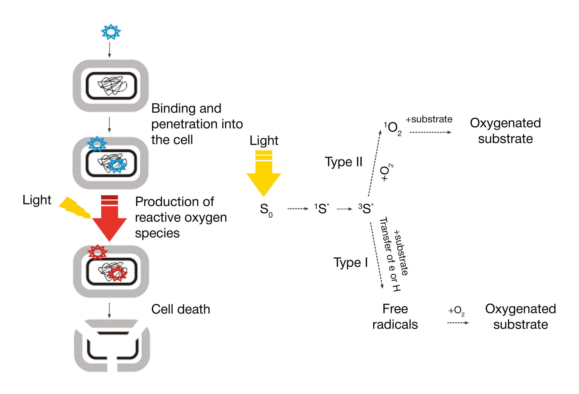
Acknowledgements: the author wishes to thank the researchers from the Laboratory of Membrane Biophysics (Department of Bioenergetics, Belozersky Institute of Physico-Chemical Biology), the Laboratory of Molecular Bioengineering (Shemyakin-Ovchinnikov Institute of Bioorganic Chemistry) and the Laboratory of Bacteriophage Genetics (Mechnikov Research Institute of Vaccines and Sera) for discussing with him some aspects of the use of bacteriophages, phage lysins and antibacterial photodynamic therapy.

Received: 2018-01-23 Accepted: 2018-01-28 Published online: 2018-03-05
|
Fig. 1. Schematic illustration of antibacterial photodynamic therapy (left). Photodynamic effects explained on the molecular level (right). Upon absorbing a photon, a photosensitizer molecule (S0) enters a short-lived excited singlet state (1S*), moving then to a triplet state (3S*). After that, two scenarios are possible: either 3S* reacts with the substrate and intermittent radicals are produced that damage cell structures and macromolecules inside the cell (type I), or energy from 3S* is transferred to oxygen, and reactive singlet oxygen 1О2 is produced that also damages cell structures or its macromolecules
Fig. 2. Possible scenarios of phage infection in the bacterial population. Reversible stages are shown in red (mutations, premature sequence termination in prophages, deletions, insertions, etc.)
Fig. 3. Schematic of bacterial cell envelopes and phage lysin classes that degrade the peptidoglycan layer