ORIGINAL RESEARCH

Clinical and molecular-genetic profiles of patients with morphological indications of congenital multicore myopathy

Kozina AA1,5, Shatalov PA3,5, Baranich TI1, Artemieva SB3, Kupriyanova AG3, Baryshnikova NV1,5, Krasnenko AYu1,5, Ilinsky VV1,2,5, Sukhorukov VS1,4
About authors

1 Pirogov Russian National Research Medical University, Moscow, Russia

2 Orekhovich Research Institute of Biomedical Chemistry, Moscow, Russia

3 Veltischev Research and Clinical Institute for Pediatrics, Pirogov Russian National Research Medical University, Moscow, Russia

4 Research Center of Neurology, Moscow, Russia

5 Genotek Ltd., Moscow, Russia

Correspondence should be addressed: Anastasia A. Kozina
Nastavnichesky per 17, bld. 1, Moscow, 105120; ur.ketoneg@rotcod

About paper

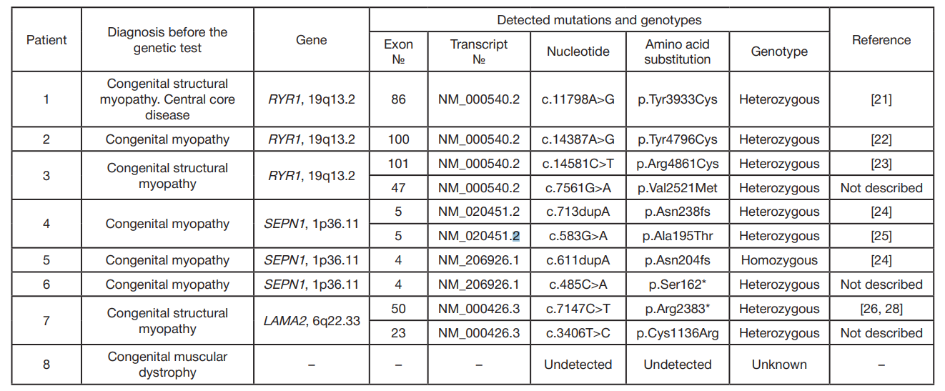
Author contribution: Kozina AA — literature analysis, analysis and interpretation of exome sequencing data, manuscript preparation; Shatalov PA — data acquisition, microscopy, manuscript preparation; Baranich TI — microscopy; Artemieva SB — medical histories and neurological examinations; Kupriyanova AG — clinical data acquisition; Baryshnikova NV — literature analysis, analysis and interpretation of exome sequencing data, manuscript preparation; Krasnenko AYu — exome sequencing; Ilinsky VV — exome sequencing; Sukhorukov VS — study design, data acquisition.

Received: 2019-02-07 Accepted: 2019-04-19 Published online: 2019-05-01
|
Fig. 1. Skeletal muscle tissue of a 21-year-old patient with congenital central core myopathy. Histochemical analysis of succinate dehydrogenase activity in frozen sections (×200; the method was proposed by Sukhorukov VS) using the nitro BT method by Nachlass et al. (1957). The total absence of enzyme activity is observed in almost all longitudinal sections of the examined muscle fibers (the arrows)
Fig. 2. Skeletal muscle tissue of a 1-year-old patient with congenital central core myopathy Histochemical analysis of succinate dehydrogenase activity in frozen sections (×200; the method was proposed by Sukhorukov VS) using the nitro BT method by Nachlass et al. (1957). The total absence of enzyme activity is observed in almost all cross-sections of the examined muscle fibers (the arrows)
Fig. 3. Skeletal muscle tissue of a 3.5-year-old patient with congenital multiminicore myopathy Histochemical analysis of succinate dehydrogenase activity in frozen sections (×200; the method was proposed by Sukhorukov VS) using the nitro BT method by Nachlass et al. (1957). Cores are abundant in almost all longitudinal sections of the examined muscle fibers (the arrows)
Table 1. Symptoms of the patients with morphological signs of core myopathies revealed by histological analysis
Note: 1 — boy, 4 years; 2 — girl, 3 years; 3 — girl, 5 years; 4 — girl, 8 years; 5 — boy, 7 years; 6 — girl, 11 years; 7 — girl, 14 years; 8 — girl, 7 years.
Table 2. A list of mutations detected in the patients with morphological signs of core myopathy
Note: 1 — boy, 4 years; 2 — girl, 3 years; 3 — girl, 5 years; 4 — girl, 8 years; 5 — boy, 7 years; 6 — girl, 11 years; 7 — girl, 14 years; 8 — girl, 7 years; * — a stop codon.