ORIGINAL RESEARCH

Bioluminescence: is it possible for a plant?

Guglya EB1, Kotlobay AA2, Sekretova EK3, Volkova PV3, Yampolsky IV1,2
About authors

1 Laboratory of Chemistry of Natural Compounds,
Pirogov Russian National Research Medical University, Moscow, Russia

2 Total Synthesis Laboratory,
Shemyakin–Ovchinnikov Institute of Bioorganic Chemistry of the Russian Academy of Sciences, Moscow, Russia

3 Moscow South-West High School No. 1543, Moscow, Russia

Correspondence should be addressed: Elena Guglya
ul. Ostrovityanova, 1, Moscow, Russia, 117997; moc.liamg@aylguge

About paper

Funding: this work was supported by the Russian Science Foundation (Grant No. 14-50-00131).

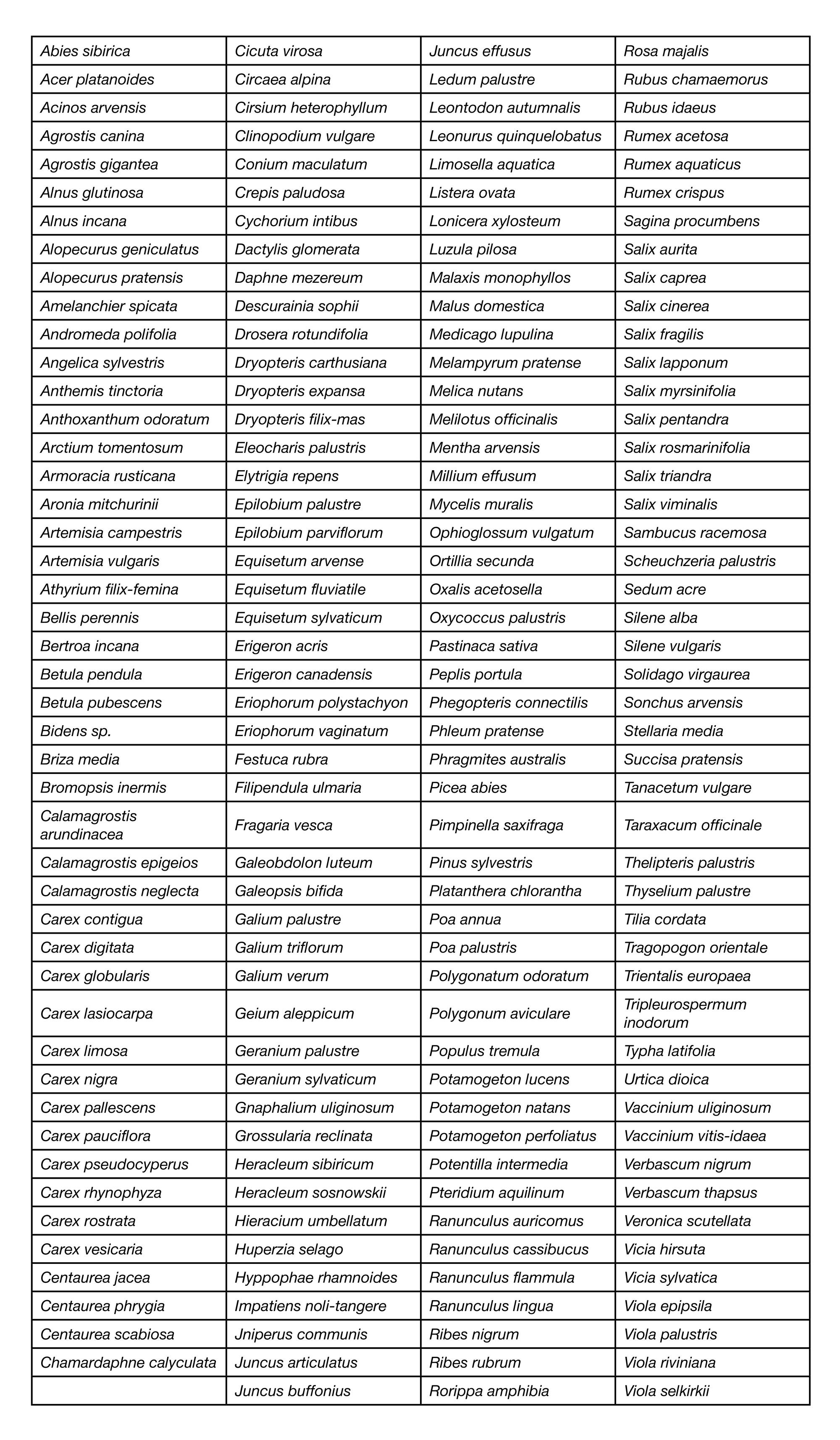
Acknowledgements: the authors thank Lyudmila Abramova and Nikita Tikhomirov for their assistance in collecting and sorting plant samples. The present research was carried out at the facilities of the Shared Resource Center of Shemyakin and Ovchinnikov Institute of Bioorganic Chemistry.

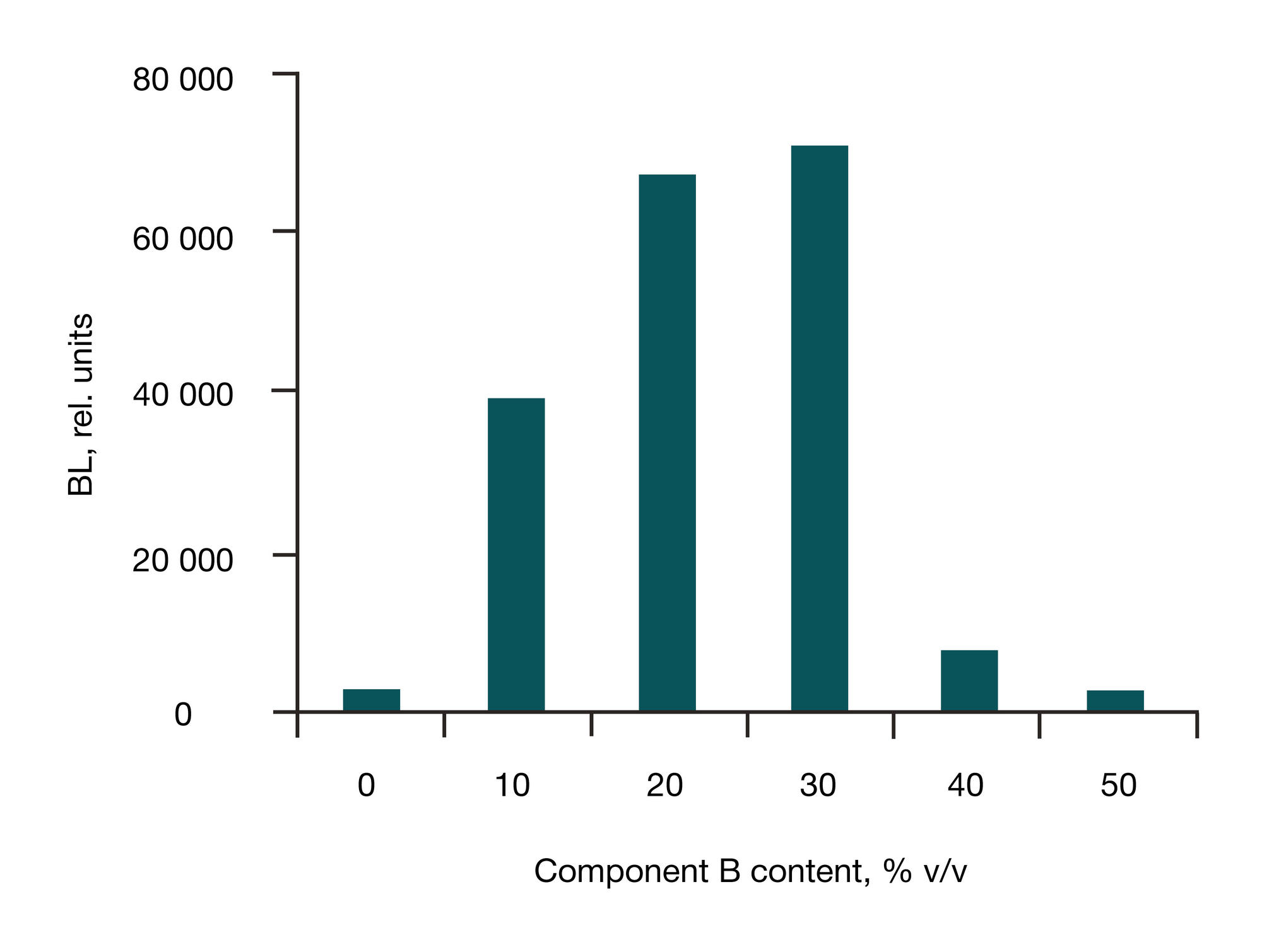
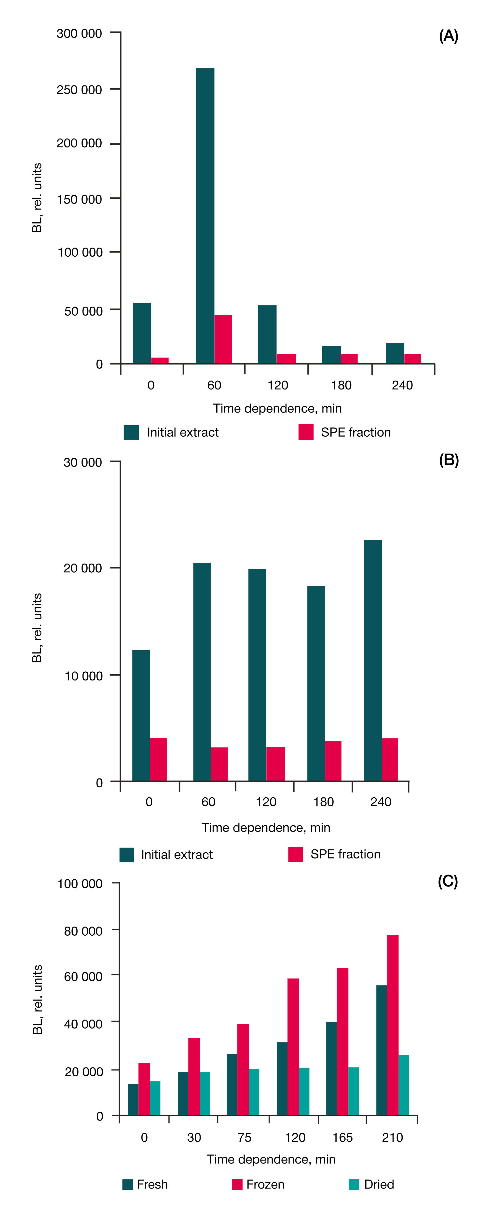
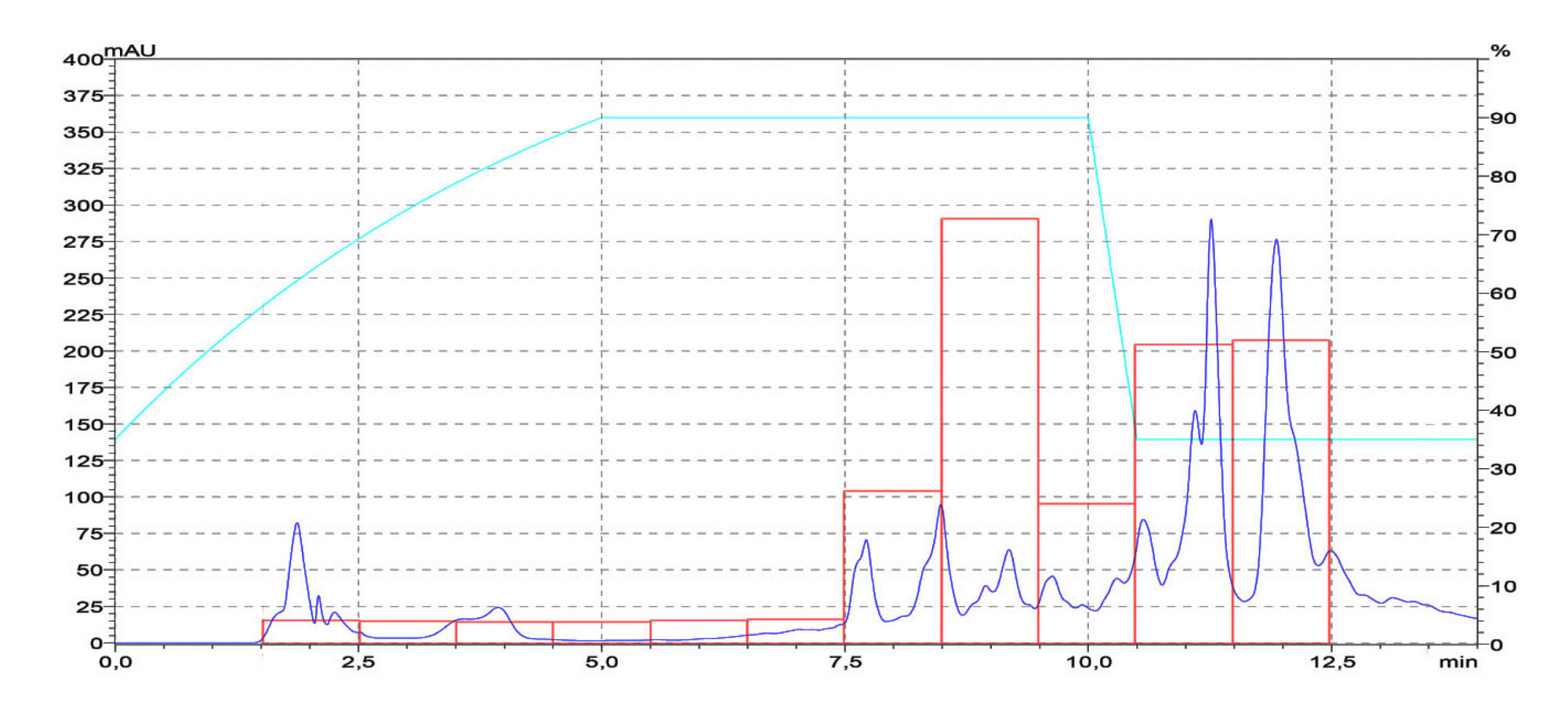
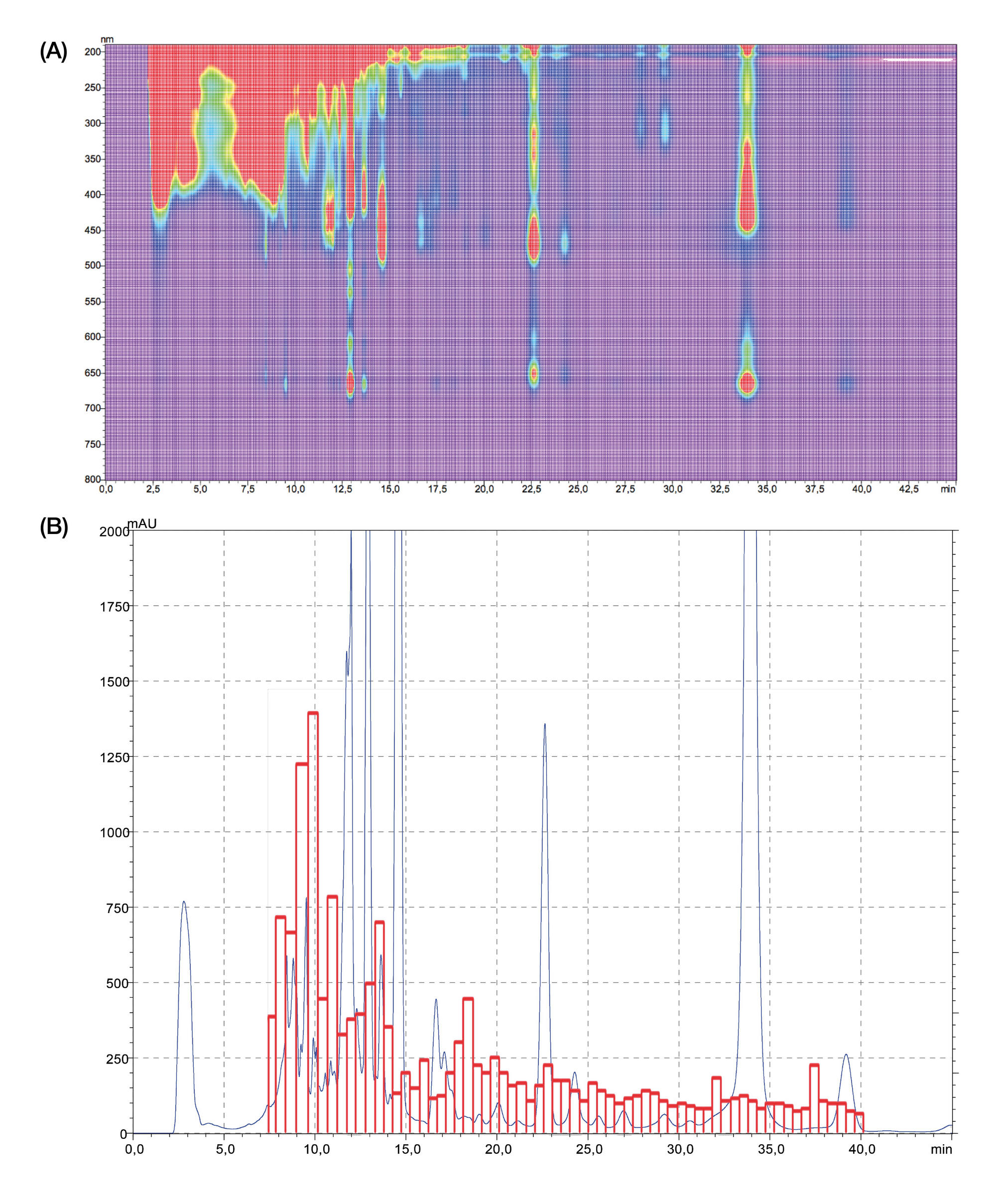
Contribution of the authors to this work: Guglya EB — preparation of organic extracts of plant samples with BL activity, development and implementation of chromatographic separations, BL analysis, drafting of a manuscript; Kotlobay AA — preparation of organic extracts of plant samples during screening of plants collection, preparation of enzyme extract, BL analysis; Sekretova EK, Volkova PV — collection and taxonomic definition of a collection of plant samples; Yampolsky IV — research planning and organization, data interpretation, editing.

Received: 2017-02-19 Accepted: 2017-04-10 Published online: 2017-06-04
|
Fig 1. Fungal bioluminescence mechanism
Fig. 2. Dependence of BL (rel. units) on component B content (% v/v) in binary solvent (A — acetone, B — 0.1 M NH4Ac, pH 6.5) for redissoving of P. natans dried extract
Fig. 3. Time dependence (min) of BL (rel. units) of leaves extracts and SPE fraction of the extracts (exposure at 0 °C): R. nigrum (A); B. pendula (B); extracts of different type material of B. pendula leaves (C)
Fig. 4. Two types of 10 s BL assay curves of B. pendula HPLC fractions
Fig. 5. HPLC separation of P. natans extract: chromatogram (UV 430 nm, blue line), luminescence of fractions (red line) and mobile phase composition (light blue line). All other conditions see Tables 2 and 3
Fig. 6. HPLC separation and luminescence of B. pendula extract: (A) — UV profile; (B) — chromatogram (UV 430 nm, blue) and activity profile (red). All conditions see Tables 2 and 3
Fig. 7. HPLC separation of P. natans extract with acetone as a mobile phase component and luminescence profile: (A) — chromatogram (UV 430 nm, blue line) and mobile phase composition (light blue line), (B) — region of 10–17 min of the chromatogram (blue line), luminescence of fractions (red line). All conditions see Tables 2 and 3
Fig. 8. Two dimensional HPLC separation of P. natans extract and luminescence profile: chromatograms (UV 430 nm, blue lines), activities profile (red lines) and mobile phase compositions (light blue line): (A) — diol column, (B) — RP column. All other conditions see in Tables 2 and 3
Table 1. Plants containing hispidin and similar compounds
Table 2. Samples for BL assay
Note. * — AB binary solvent (A — acetone, B — 0.1 M NH4Ac, pH 6.5).
Table 3. HPLC conditions
Note. * — Component A — 0.02 M NH4Ac, pH 5.5.
Table 4. BL activity of plant extracts
Table 5. Losses of activity in separation process (see Fig. 8)
Note. * — V1 — total solution volume; V2 — solution volume in measurement; ** — A = a · V1/V2.
Table 1S. Plant species of Central Russia tested for a substrate of bioluminescent reaction