METHOD

Multimodal optical coherence tomography in the assessment of cancer treatment efficacy

Sirotkina MA1, Kiseleva EB1, Gubarkova EV1, Buyanova NL1, Elagin VV1, Zaitsev VYu1,2, Matveev LA1,2, Matveev AL1,2, Kirillin MYu2, Gelikonov GV1,2, Gelikonov VM1,2, Kuznetsov SS1, Zagaynova EV1, Gladkova ND1
About authors

1 Department of Microbiology and Immunology,
Nizhny Novgorod State Medical Academy, Nizhny Novgorod, Russia

2 Institute of Applied Physics, Russian Academy of Sciences, Nizhny Novgorod, Russia

Correspondence should be addressed: Marina Sirotkina
pl. Minina i Pozharskogo, d. 10/1, Nizhny Novgorod, Russia, 603950; ur.liam@m_aniktoris

About paper

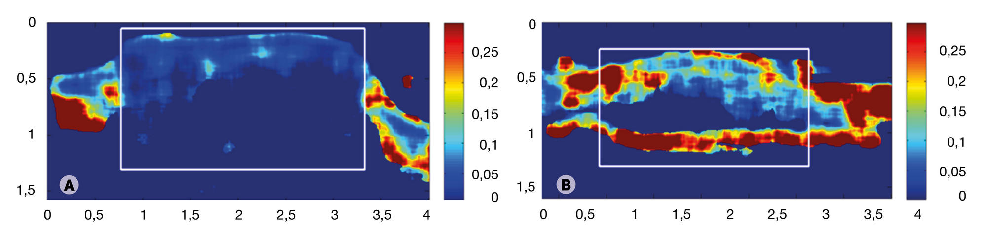
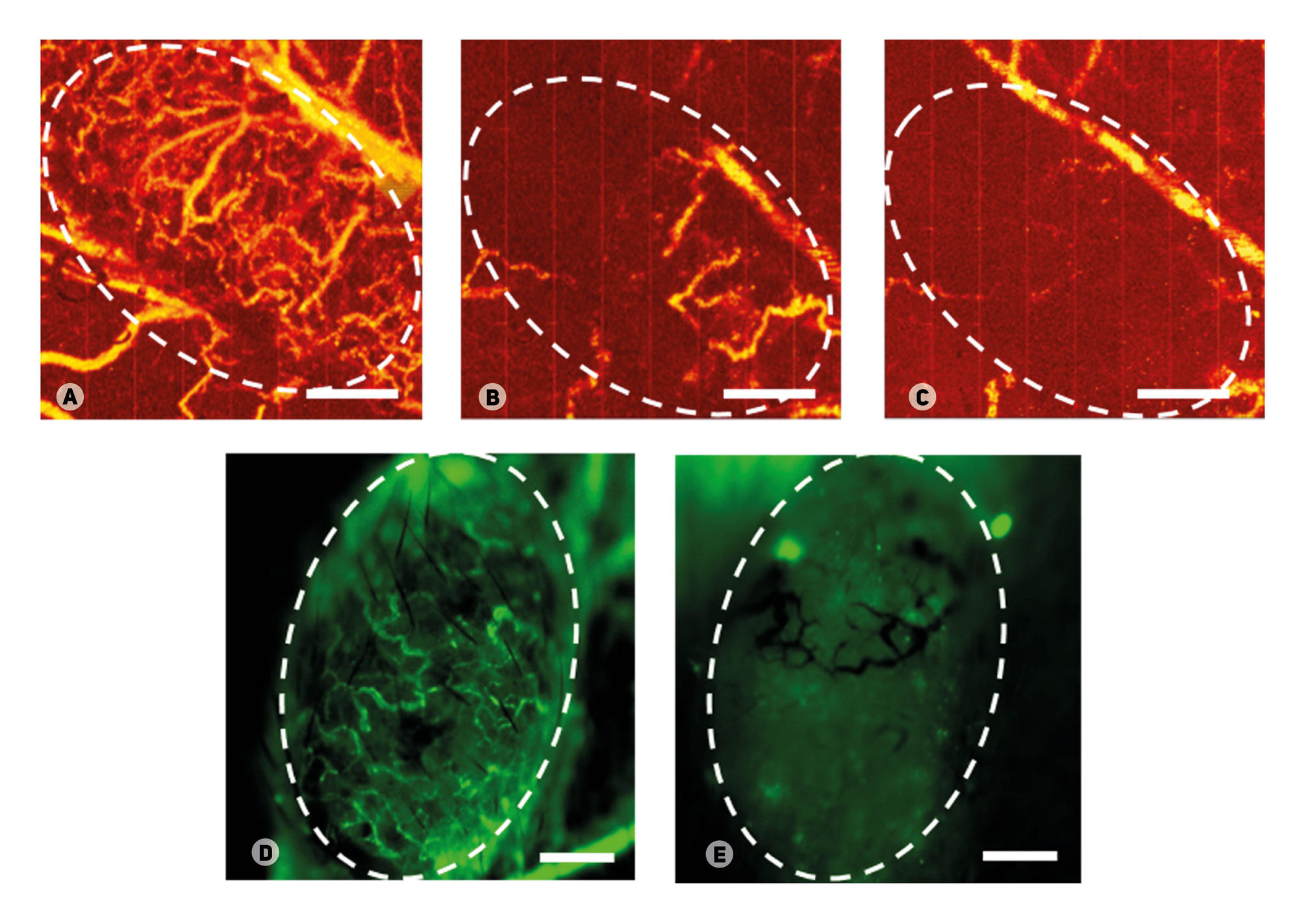
Funding: this work was supported by the Ministry of Education and Science of the Russian Federation (grant no. 14.B25.31.0015), numerical processing of CP OCT images was supported by grant no. 15-32-20250 of the Russian Foundation for Basic Research, algorithm modification and development of software/ hardware OCT-system for mapping microcirculation and elastography images was supported by the grant of the President of the Russian Federation for young scientists (no. MK-6504.2016.2) and the grant of the Russian Foundation for Basic Research (no. 16-02-00642-а).

Acknowledgements: authors thank professor Alex Vitkin of University of Toronto (Toronto, Canada), the leading scientist of the Russian Federation Government Mega-grant 14.B25.31.0015.

Received: 2016-08-15 Accepted: 2016-08-25 Published online: 2017-01-05
|
Fig. 1. Visualization of healthy tissue (A, B) and CT26 colon carcinoma transplanted into the mouse ear (C, D)
(A) and (C) are histological sections (the scale bars are 100 μm and 500 μm, respectively). (B) and (D) are images obtained with cross-polarized optical coherence tomography (the scale bar is 1 μm). 1 — the cartilage; 2 — the adipocyte layer; 3 — the cross-striated muscle; 4 — the dermis; 5 — hair follicles; 6 — the epidermis; 7 — the tumor tissue.
Fig. 2. Visualization of a vascular network of healthy tissue (A–C) and CT26 colon carcinoma transplanted into the mouse ear (D–F)
(A) and (B) are microphotographs, (C) and (D) are images obtained with OCT-based microangiography, (E) and (F) are fluorescence microscopy images. The scale bar is 0.5 mm.
Fig. 3. Structured in vivo OCT images (A, C) and elastographic deformation maps (B, D) of healthy tissues of the mouse ear (A, B) and CT26 colon carcinoma transplanted into the mouse ear (C, D), in vivo (the scale bar is 1 mm)
Fig. 4. MM OCT-based monitoring of CT26 colon carcinoma transplanted into the mouse ear
Top row: CP OCT images; bottom row: OCT MA images. The tumor site is circled.
Fig. 5. Depolarization maps based on B-scans. (A) — tumor before photodynamic therapy, (B) — tumor after photodynamic therapy
Fig. 6. Visualization of the vascular response of CT26 colon carcinoma transplanted into the mouse ear to photodynamic therapy on OCT MA (A–C) and fluorescence microscopy (D, E). (A) and (D) — before treatment, (B) — immediately after treatment, (C) and (E) — 24 h after treatment. The scale bar is 0.5 mm. The tumor site is marked with a dotted circle