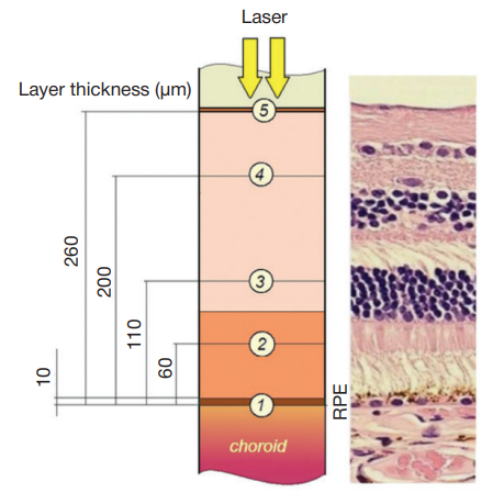
Fig. 1. A topographic representation of the chorioretinal complex.
1— the retinal pigment epithelium (10 μm in thickness), 2 — the layer of photoreceptors (60 μm), 3 — the outer nuclear layer (110 μm), 4 — the inner nuclear layer (200 μm), 5 — the inner limiting membrane (260 μm)
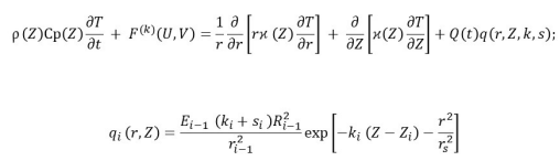
Fig. 2. The heat equation for layer i of the model.
T (r, Z, t) is a temperature rise above the physiological norm (37 °С); Cp (Z), p (Z), ϰ (Z), are specific heat capacity, density and thermal conductivity, respectively; Q(t) is time-dependent intensity of the absorbed radiation; q is volumetric heat generation, k and s are light absorption and scattering; F(k) is a function determined by the rates of radial (V) and axial (U) convective heat transfer; Ri-1 and Ei-1 are spot size (eхp (-1)) and Gaussian beam irradiance at the interface between two adjacent layers
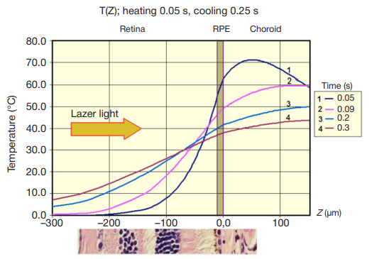
Fig. 3. A relationship between the degree of retinal tissue heating and the
thickness Z (r = 0) of the chorioretinal complex by the end of the pulse, t = 0.05 s (1) and during tissue cooling 0.09 s (2), 0.2 s (3) and 0.3 s (4) after the onset of irradiation
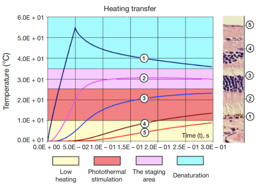
Fig. 4. Heating and cooling dynamics T (t, Zi, r = 0) in the retinal zones corresponding to the thickness Zi of layer
1 (the retinal pigment epithelium, 10 μm), 2 (the layer of photoreceptors, 60 μm), 3 (the outer nuclear layer, 110 μm), 4 (the inner nuclear layer, 200 μm), and 5 (the inner limiting membrane, 260 μm). The blue color marks the area of irreversible protein denaturation at temperatures that exceed the physiological norm by 25 °С. The area of photothermal stimulation with the temperature rise ranging between 10 and 25 °С is shown in red; at these temperatures the therapeutic effect is achieved, and irreversible protein denaturation is almost negligible. The staging area marked in purple is where tissue regeneration stops and irreversible protein denaturation begins
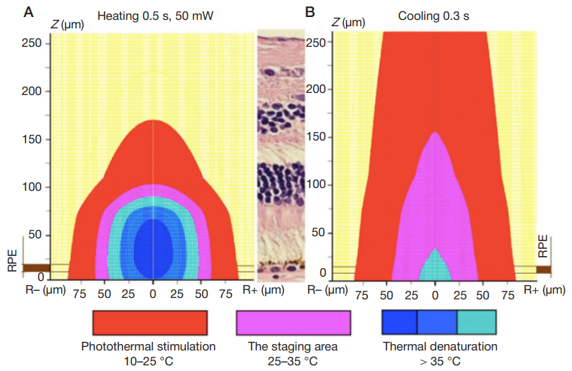
Fig. 5. Distribution of temperatures T (r, Z) in retinal tissues during heating (А) and cooling (B) by the end of the laser pulse (t = 0.05 s) and at t = 0.3 s (cooling) in the sagittal plane
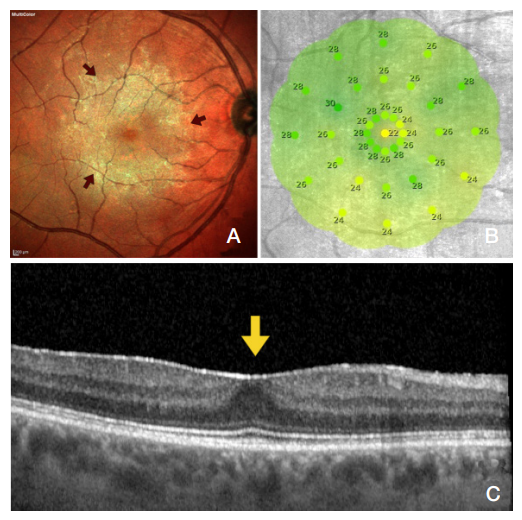
Fig. 6. The ocular fundus before the treatment
(A) a Multicolor photo showing the surface and the boundaries of ERM in yellow and green (marked by red arrows); (B) microperimetry of the macular zone, the average light sensitivity was 26.3 dB; (C) SOCT of the retinal membrane surface showing a hyperreflective line fused to the inner limiting membrane; the foveal pit is flat (the yellow arrow); the thickness of the central retina is 257 μm
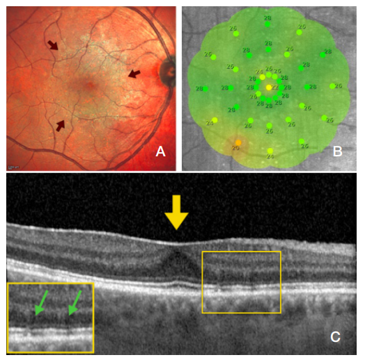
Fig. 7. The ocular fundus after the first session of grid laser photocoagulation
(A) a Multicolor photo of the fundus shows thinner, blurred ERM contours in the macular zone (the red arrow); (B) microperimetry of the macular zone: the average light sensitivity is 26.5 Db; (C) SOCT reveals a hyperreflective line fused to ILM, the foveal pit is flat (the yellow arrow), the thickness of the central retina is slightly decreased (253 μm). The green arrow on the OCT image marks laser coagulates; the retinal layers are structurally unchanged
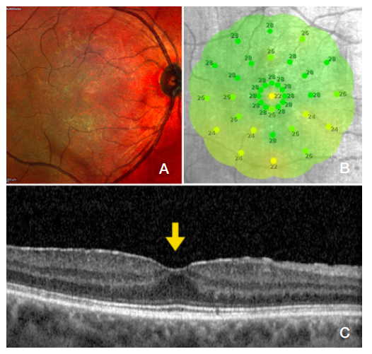
Fig. 8. The ocular fundus after the completed treatment course: (A) a Multicolor photo of the fundus showing ERM regression; (B) microperimetry: the average light sensitivity is 26.6 dB; (C) an OCT image showing a thinner hyperreflective line (ERM) and formation of the foveal pit (the yellow arrow); the thickness of the central retina is 246 µm
